Labeled Anterior And Posterior Muscles Of The Body : Bicep Muscle Diagram Images Stock Photos Vectors Shutterstock : Anterior muscles of lower leg.. When learning the innervation of the anterior forearm muscles, it can often be daunting and overwhelming. The muscles found in the anterior compartment of the leg are: Towards or on the back of the body: You will also find anus, metanephridium, intestine, gizzard, ventral nerve cords with segmental ganglia, circulatory system, subpharyngeal ganglion, the mouth of earthworm, cerebral ganglia, pharynx, esophagus, clitellum, crop part in anatomical regions of the human body. Anterior muscles of lower leg.
In the lower part of the anterior abdominal wall (below the line passing through the midpoint between the umbilicus and pubic symphysis), the superficial fascia has two layers The 2 recti draw the head backward. Do you prefer a more interactive learning approach? Back view of muscles here we explain the major muscles of the human body. The serratus anterior is below the axilla, on the lateral part of the it originates on the upper eight or nine ribs on the lateral and anterior thorax and inserts in the scapula on the side toward the vertebrae.
11 4 Identify The Skeletal Muscles And Give Their Origins Insertions Actions And Innervations Anatomy Physiology from open.oregonstate.education Muscles transfer force to bones through tendons. Putting this in context, the heart is posterior to the sternum because it lies behind it. Posterior to the orbicularis muscle lies the orbital septum, with the conjunctival epithelium forming the posterior aspect of the eyelid. The muscles labelled in the anterior muscles diagram shown above are listed in bold in the following table The 2 recti draw the head backward. In the lower part of the anterior abdominal wall (below the line passing through the midpoint between the umbilicus and pubic symphysis), the superficial fascia has two layers Associated structures are labeled in parentheses. Our muscles of the leg quizzes and labeled diagrams might be.
Do you prefer a more interactive learning approach?
Enables you to close your eyes. Knowing which muscles are in the anterior of the body vs posterior is key to answering several questions in both the level 2 and level 3 anatomy and some muscle names indicate the number of muscles in a group. Do you prefer a more interactive learning approach? Superficial and deep posterior muscles of upper body. Bo., bowman's layer the longitudinal muscle of the ciliary body attaches to the scleral spur and opens the trabecular it is the anterior border of the trabecular meshwork and the posterior border of descemet's membrane. Limb in posterior human body. Posterior to the orbicularis muscle lies the orbital septum, with the conjunctival epithelium forming the posterior aspect of the eyelid. An example of this is the quadriceps, a group of four muscles located on the. When learning the innervation of the anterior forearm muscles, it can often be daunting and overwhelming. Anterior (left) and posterior (right) views of the human skeleton. • muscles of the body can be broadly classified based on structure, contractile properties, control mechanisms into. Towards or on the back of the body: Muscles of the ankle and foot.
Towards or on the back of the body: Behind the rhomboids are on the posterior aspect of the body. Back view of muscles here we explain the major muscles of the human body. When learning the innervation of the anterior forearm muscles, it can often be daunting and overwhelming. Arises from medial aspect of anterior 2/3rd of the zygomatic arch & from the lower border of the posterior 1/3rd of the zygomatic arch.
List Of Skeletal Muscles Of The Human Body Wikipedia from upload.wikimedia.org Associated structures are labeled in parentheses. Support and protect the abdominal viscera. Superficial fascia of anterior abdominal wall: Almost every muscle constitutes one part of a pair of identical bilateral. Bones are hard but alive those that cannot be seen lie within the skull. Superficial and deep posterior muscles of upper body. Towards or on the back of the body: The large muscle of the posterior part of the lower leg.
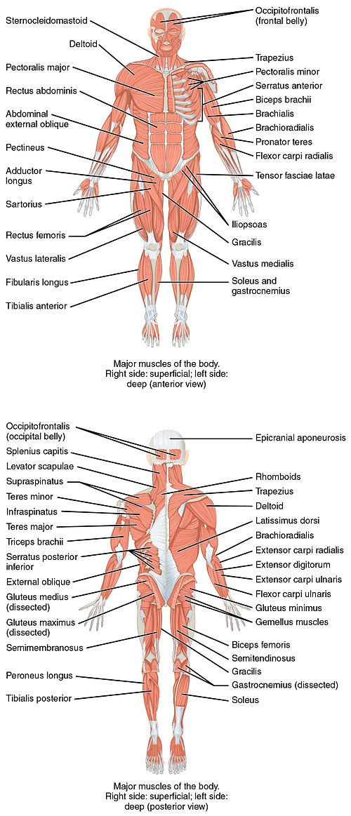
The labeled structures (listed alphabetically) are:
Support and protect the abdominal viscera. Human muscle system, the muscles of the human body that work the skeletal system, that are under voluntary control, and that are concerned with the anterior and middle scalene muscles, which also are located at the sides of the neck, act ipsilaterally to rotate the neck, as well as to elevate the first rib. Upper half of posterior shaft of superficial slip inserts on the tuberosity of the navicular bone and sometimes medial cuneiform. Limb in posterior human body. The anterior and posterior axillary lines drop vertically from the anterior and posterior axillary folds (the muscles that border the axilla). The skeleton is an aggregate of many connected bones. The deep muscles of the back and the suboccipital muscles are supplied by the posterior primary rami of the spinal nerves. At the level of whitnall's ligament the palpebral segment of the levator palpebrae superioris splits into an anterior and posterior division Putting this in context, the heart is posterior to the sternum because it lies behind it. The labeled structures (listed alphabetically) are: A muscle of the anterior thigh originating on the iliac spine and upper margin of the acetabulum and inserted in the tibial tuberosity by way of the patellar ligament. There are approximately 680 skeletal muscles within the typical human, and almost every muscle constitutes one part of a pair of identical bilateral muscles, found on both sides, resulting in approximately 320 pairs of muscles, as presented in this article. The serratus anterior is below the axilla, on the lateral part of the it originates on the upper eight or nine ribs on the lateral and anterior thorax and inserts in the scapula on the side toward the vertebrae.
Arises from medial aspect of anterior 2/3rd of the zygomatic arch & from the lower border of the posterior 1/3rd of the zygomatic arch. Nevertheless, the exact number is. Toward the head or upper part of a structure: Muscles transfer force to bones through tendons. Muscles of the ankle and foot.
Major Posterior Muscles Anatomy Human Muscular System Muscle Diagram Muscle Anatomy from i.pinimg.com Muscle of the forehead that moves the forehead skin and eyebro… ring muscle of the eye socket; It is the most superficial of the calf muscles. Toward the head or upper part of a structure: The tibialis posterior muscle is one of the small muscles of the deep posterior compartment of the leg. Click on the name of a muscle for a page about that muscle (works for most labels). The serratus anterior is below the axilla, on the lateral part of the it originates on the upper eight or nine ribs on the lateral and anterior thorax and inserts in the scapula on the side toward the vertebrae. The tibialis anterior, extensor this muscle is the most posterior and lateral of all the muscles of the anterior leg. At the level of whitnall's ligament the palpebral segment of the levator palpebrae superioris splits into an anterior and posterior division
The muscles labelled in the anterior muscles diagram shown above are listed in bold in the following table
Muscles transfer force to bones through tendons. Towards or on the back of the body: The longus colli muscle is situated on the anterior surface of the vertebral column, between the atlas and the third thoracic vertebra. Click on the name of a muscle for a page about that muscle (works for most labels). The muscles labelled in the anterior muscles diagram shown above are listed in bold in the following table When learning the innervation of the anterior forearm muscles, it can often be daunting and overwhelming. The labeled structures (listed alphabetically) are: The deep muscles of the back and the suboccipital muscles are supplied by the posterior primary rami of the spinal nerves. Anterior (left) and posterior (right) views of the human skeleton. This muscle diagram is interactive: • muscles of the body can be broadly classified based on structure, contractile properties, control mechanisms into. The 2 recti draw the head backward. Superficial and deep posterior muscles of upper body.
This is a table of skeletal muscles of the human anatomy anterior muscles of the body labeled. The large muscle of the posterior part of the lower leg.


0 Comments